Virtual ToolBox
Here you will find research tools available within EuroCurvoBioNet to study curvature -biology interactions at the level of biological membranes, single cells and cell collectives in curved environments. A particular focus is given on time resolved 3D imaging and the associated image analysis to study single cells in curved environments. Mathematical tools are also available to explore the role of curvature at multiple length scales with a focus on open-source implementations.
Experimental
Nanometre, Micrometre, Millimetre, Centimetre
Manufacturing
3D-printing equipment down to sub-µm resolution
Light-based 3D printing equipment which can create curved 3D substrates based on biocompatible and/or cell-interactive polymers (in a wide range of resolutions ranging from sub-µm to µm). The constructs can have dimensions up to cm³ scale.

Relevant links:
Contact:
Sandra Van Vlierberghe
Ghent University
Experimental
Nanometre, Micrometre, Millimetre, Centimetre
Manufacturing
UpNano and Nanoscribe Multiphoton 3D printers
Multiphoton polymerisation is the highest resolution additive manufacturing technique, capable of printing features down to ~200nm. At Nottingham, we have both a Nanoscribe GT2 and an UpNano NanoOne. Our Nanoscribe is also fitted with the Heteromerge module, allowing multi-material 3D printing. The UpNano is capable of printing in a sterile, heated, humidified environment suitable for live cells.

Relevant links:
Contact:
Robert Owen
University of Nottingham
Experimental
Nanometre, Micrometre
Imaging
Deformable hydrogel microparticles
Microparticles that serve as cellular force sensors. Their rigidity (0.1 - 50 kPa) and size (3 - 25 um dimater) can be controlled. Their deformations can be determined with a <100 nm resolution after conventional confocal imaging. Especially useful for studying cell-cell interactions, and cellular machinery involved in generating forces. Can also be embedded or microinjected into multicellular tissue.

Relevant links:
Contact:
Daan Vorselen
Wageningen University
Experimental
Nanometre, Micrometre
Manufacturing
Nanoscribe GT 2
2 Photon Polymerization printer for structures in the micrometer to submicrometer scale.

Relevant links:
Contact:
Barbara Schamberger
Heidelberg University
Experimental
Nanometre, Micrometre, Millimetre, Centimetre
Imaging
Nanoscale and Microscale ResearchCentre
The NMRC is an inter-disciplinary facility dedicated to supporting and promoting world-leading nanoscience and materials characterisation.
Facilities include:
Scanning Electron Microscopy (SEM), Transmission Electron Microscopy (TEM), Atomic Force Microscopy (AFM), Raman Spectroscopy, Secondary Ion Mass Spectrometry (SIMS), X-ray Photoelectron Spectroscopy (XPS), Nanofabrication Suite (ebeam lithography), Biophysical Analysis, Cryogenic Materials Characterisation, Particle Sizing, Confocal Laser Scanning Microscopy, and a dedicated suite for CL2 cell culture.

Relevant links:
Contact:
Robert Owen
University of Nottingham
Experimental
Micrometre
Analysis
Fast-Fourier Transform Electrochemical Impedance spectroscopy
Allows for the measurement of the electric properties (resistance and capacitance) of planar lipid bilayers

Relevant links:
Contact:
Ognyan Petkov
Institute of Solid State Physics, Bulgarian Academy of Sciences
Experimental
Micrometre
Manufacturing
2D and 3D protein patterning
A method to generate protein patterns on (flat or curved) surfaces.

Relevant links:
Contact:
Nicholas Kurniawan
Eindhoven University of Technology
Experimental
Micrometre
Analysis
Thermal shape fluctuation analysis (TSFA) of giant unilamellar vesicles (GUV)
The method for the bending elasticity measurement of lipid membranes via thermal shape fluctuation analysis (TSFA) is based on the autocorrelation analysis of thermal shape fluctuations of quasispherical giant (~10-100 micrometers in diameter) unilamellar vesicles (GUV). The TSFA exploits the relation between the bending elasticity of the lipid bilayer, its membrane tension and the amplitudes of GUV's shape fluctuations, due to the Brownian motion of the water molecules, surrounding the membrane. The non-invasive approach consists in videomicroscopy of the fluctuating GUV membrane, followed by digitization of vesicle images and mathematical analysis of the recorded fluctuations, yielding the bending modulus of the membrane with high accuracy (~ 5%) due to the stroboscopic illumination of the sample.

Relevant links:
Contact:
Victoria Vitkova
Institute of Solid State Physics, Bulgarian Academy of Sciences
Experimental
Micrometre
Manufacturing
Digital hydrogel photosculpting
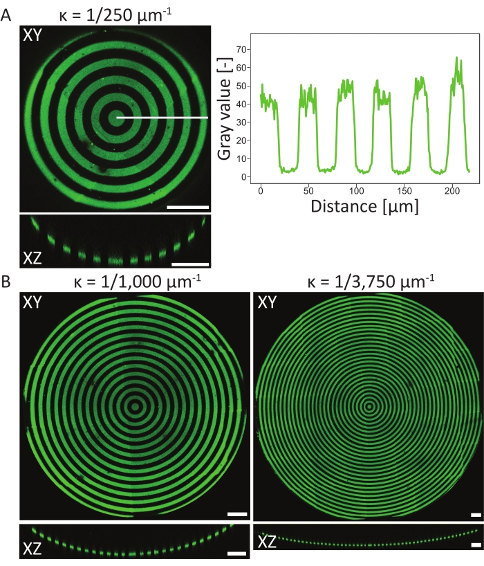
We developed a light-based method to create stable microscale topographies on diverse types of hydrogels (including polyacrylamide, GelMA, etc). Gradients and complex-shape topographies can also be robustly created.

Relevant links:
Contact:
Nicholas Kurniawan
Eindhoven University of Technology
Experimental
Micrometre
Manufacturing
Hydrogel with on-demand topographies
We developed a method to non-invasively create and erase microscale topograhies on cell-compatible photoresponsive hydrogel.

Relevant links:
Contact:
Nicholas Kurniawan
Eindhoven University of Technology
Experimental
Micrometre
Manufacturing
Microstructures from 3D models using grayscale lithography
We developed a method to create microstructured cell culture substrates from 3D models using grayscale lithography

Relevant links:
Contact:
Laurent Pieuchot
CNRS
Experimental
Micrometre, Millimetre
Imaging
Microfluidics, advanced imaging and analysis, polarization optics, AFM, photophysiology (PAM), ML-based algorithms, CFD
The description of the relevant instruments listed above could be found in published literature form my lab.

Relevant links:
Contact:
Anupam Sengupta
University of Luxembourg
Experimental
Micrometre, Millimetre
Manufacturing
Curvo-chip
A microfabricated PDMS chip containing a library of curved structures (convex and concave cylinders, spheres, torus).

Relevant links:
Contact:
Nicholas Kurniawan
Eindhoven University of Technology
Experimental
Micrometre, Millimetre
Manufacturing
Fluidic chip with adaptable sizes channels and gels
A fluidic chip that allows for template casting channels in custom gels. Allows for apical and basolateral sampling, fluorescent imaging and 4 channels run parallel both flow or left staticly.

Relevant links:
Contact:
Ronald van Gaal
Utrecht University
Experimental
Micrometre, Millimetre
Manufacturing
Projection Microstereolithography 3D Printers
Projection microstereolithograohy is a layer-by-layer 3D printing technique. Compared to 2PP/MPL, it is much faster at manufacturing, but at lower resolution. We have two Boston Microfabrication (BMF) systems, the S130 (2um pixel size) and the S240 (10um pixel size). These systems are designed for industrial manufacturing and can achieve scale versus smaller systems. The S240 is also housed within a microbiological safety cabinet for live cell printing.

Relevant links:
Contact:
Robert Owen
University of Nottingham
Experimental
Micrometre, Millimetre
Imaging
3D scanning and 3D printing
We obtain CAD data by scanning surfaces with the 3D optical scanning system in our laboratory. Then, we can physically produce three-dimensional prototype models from this data by using 3D printers. We can perform dimensional, geometric and surface analysis of the models we produce.

Relevant links:
Contact:
Binnur Sagbas
Yildiz Technical University
Experimental
Micrometre, Millimetre
Manufacturing
Bioprinter
Bioprinter enables the production of designed three dimensional structures using cell-laden soft polymers (also known as bioinks typically hydrogels+cells). The structure of the bioprinted object can be digitally designed and given the use of rheologically and structurally competent bioinks. Each filament extruded can be 100-200 micrometers in diameter and mm - cm structures can be typically produced. Besides extrusion bioprinting we also offer laser assisted bioprinting which can print droplets of 10-100 micrometers containing cells. This technology can generate larger tissue structures bottom up in additive manufacturing mode of operation.

Relevant links:
Contact:
Ioannis Papantoniou
KULeuven
Experimental
Micrometre, Millimetre
Imaging
3D Lightsheet Microscpy
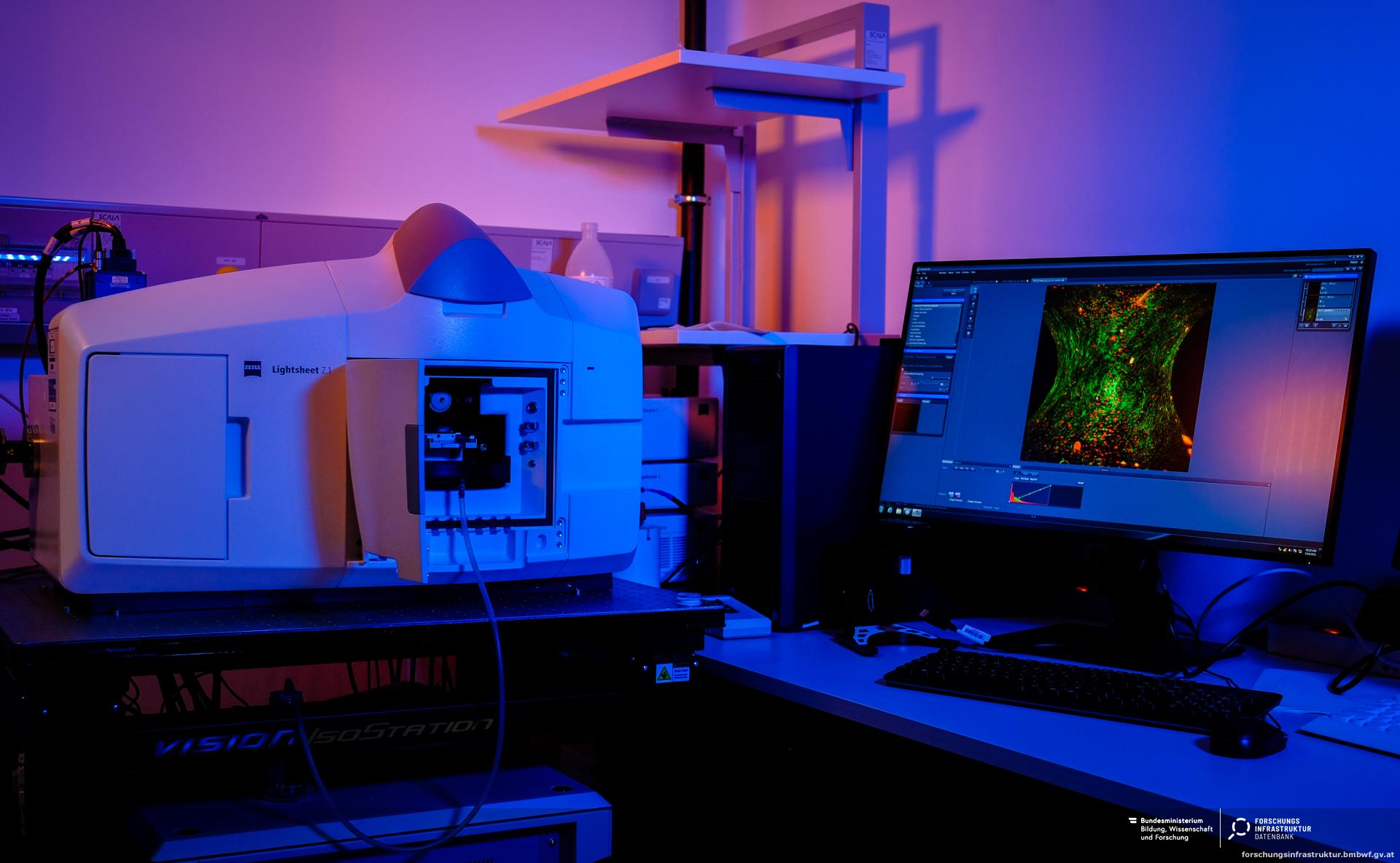
Microscope ZEISS Lightsheet Z1
Light Sheet Microscopy allows measurements of 3D Cell Cultures and tissues. The measurements can be performed on fixed and living cell cultures. The sample chamber is set up to allow for controlled conditions of humidity, temperature and CO2. A range of laser sources are available (405nm, 488nm, 561nm, and 638nm) to allow for standard measurements of typical fluorophores. 10x and 20x water objectives are available.
The system also allows measuring of cleared samples after refractive index matching with a 20x clearing objective and the respective sample chamber.

Relevant links:
Contact:
Andreas Roschger
Paris Lodron University of Salzburg
Experimental
Micrometre, Millimetre
Manufacturing
Static and dynamic curved substrates
Deformable substrates controlled by magnetic actuation and micromachining of curved substrates

Relevant links:
Contact:
Caterina Tomba
Institute of Nanotechnologies of Lyon (INL, CNRS)
Experimental
Micrometre, Millimetre, Centimetre
Imaging
Multiphoton Laser Scanning Microscope (Evident)
Confocal and Multiphoton laser scanning microscope with a resonance scanner for fast imaging

Relevant links:
Contact:
Barbara Schamberger
Heidelberg University
Experimental
Millimetre, Centimetre
Functional
Electrophysiology, 3D cultures
Electrophysiological study of membranes and assessment of transmembrane resistance and ionic currents.

Relevant links:
Contact:
Sotirios Zarogiannis
Faculty of Medicine, School of Sciences, University of Thessaly
Experimental
Millimetre, Centimetre
Manufacturing
Bioreactor to culture tubular scaffolds and tissues
A bioreactor that can be used to culture tubular constructs with controlled and independent application of shear flow and stretch.

Relevant links:
Contact:
Nicholas Kurniawan
Eindhoven University of Technology
Computational
Nanometre
Modelling
SPIRE - software tool for bicontinuous phase recognition
An interactive tool for recognizing bicontinuous structures in TEM sections based on comparison to mathematical "nodal surface" models.

Relevant links:
Contact:
Łucja Kowalewska
University of Warsaw
Computational
Nanometre
Modelling
Differential geometry
I use the methods of differential geometry in the consideration of curves and surfaces. In particular, I study curvature-based functions and their variations during infinitesimal bending .

Relevant links:
Contact:
Marija Najdanović
University of Priština in Kosovska Mitrovica, Faculty of Sciences and Mathematics, Serbia
Computational
Nanometre, Micrometre
Imaging
BiaPy
BiaPy is an open source ready-to-use all-in-one library that provides deep-learning workflows for a large variety of bioimage analysis tasks, including 2D and 3D semantic segmentation, instance segmentation, object detection, image denoising, single image super-resolution, self-supervised learning, image classification and image-to-image translation.

Relevant links:
Contact:
Ignacio Arganda-Carreras
University of the Basque Country (UPV/EHU)
Computational
Nanometre, Micrometre, Millimetre, Centimetre, Metre and larger
Analysis
Computational implementation of curvature estimation on triangle meshes
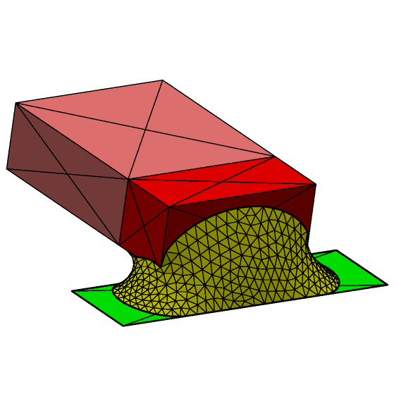
Python codes for taking an stl file and calculating local curvatures, using libigl-based implementations. Simple to use, and easy to visualise in VTK (VTK files are automatically generated). Also possible to create interface shape distributions (k1k2 plots), and generate the input files for Karambola (software for Minkowski functional calculation).

Relevant links:
Contact:
Sebastien Callens
Eindhoven University of Technology
Computational
Nanometre, Micrometre, Millimetre, Centimetre, Metre
Modelling
Surface Evolver
A tool developed by Kenneth Brakke to simulate energy minimisation of surfaces. (Ideally we should try to create a list of "power users" who know how to do various tricks in the software and are willing to help and collaborate).

Relevant links:
Contact:
John Dunlop
University of Salzburg
Computational
Micrometre
Modelling
Quantification of stress fibers and focal adhesion
An image-based algorithm to quantify morphology of stress fibers and focal adhesions from microscopy images of cells.

Relevant links:
Contact:
Nicholas Kurniawan
Eindhoven University of Technology
Computational
Micrometre
Modelling
Lumerical FDTD
3D Simulations of the optical response of a given nanostructure

Relevant links:
Contact:
Bodo Wilts
University of Salzburg
Computational
Micrometre
Modelling
Numerical computation of continuous equations
I use continuous equations based on physical principles: conservation laws (hydrodynamics), electromagnetism, phase transition to capture dynamics in soft materials such as lipid membranes and polyelectrolyte gels that may be relevant for biological functions. To give one example, I use these tools to unravel the role of nonelectric aspects of action potentials in information processing.

Relevant links:
Contact:
Matan Mussel
University of Haifa
Computational
Micrometre, Millimetre
Modelling
Tissue Simulation Software
A software tool able to implement simulations of single or multi-component epithelial tissues including gene regulation, mechanical properties of cells, and mechanobiology feedbacks.

Relevant links:
Contact:
Javier Buceta
Institute for Integrative Systems Biology
Computational
Micrometre, Millimetre
Imaging
CartoCell
We introduce CartoCell, a deep-learning-based pipeline that uses small datasets to generate accurate labels for hundreds of whole 3D epithelial cysts. Our method detects the realistic morphology of epithelial cells and their contacts in the 3D structure of the tissue. CartoCell enables the quantification of geometric and packing features at the cellular level. Our single-cell cartography approach then maps the distribution of these features on 2D plots and 3D surface maps, revealing cell morphology patterns in epithelial cysts. Additionally, we show that CartoCell can be adapted to other types of epithelial tissues.

Relevant links:
Contact:
Luis M. Escudero
Seville University
Computational
Micrometre, Millimetre
Modelling
Agent-Based Modelling
open-source simulation platform for agent-based modelling of biological systems

Relevant links:
Contact:
Vasileios Vavourakis
University of Cyprus
Computational
Micrometre, Millimetre, Centimetre
Modelling
BioDynaMo
BioDynaMo is a high-performance software for the computational modelling of tissue development. It is an agent-based modelling software, so the user can specify behaviours that individual cellular elements follow, in order to model collective cellular behaviour, including tissue dynamics. In particular, one can simulate biomechanics and biological behaviours, such as for instance the development of curvature.

Relevant links:
Contact:
Roman Bauer
University of Surrey, UK
Computational
Micrometre, Millimetre, Centimetre
Modelling
Finite Element Modelling Procedures
suite of open-source solvers and simulation tools relevant to biomechanical modelling

Relevant links:
Contact:
Vasileios Vavourakis
University of Cyprus
Computational
Micrometre, Millimetre, Centimetre, Metre and larger
Modelling
Finite element analysis
Finite element analysis is a numerical method used to approximate solutions to complex engineering problems by breaking down structures into smaller elements. It is particularly powerful in Multiphysics applications, where it can simultaneously solve coupled physical phenomena such as diffusion and structural interaction.

Relevant links:
Contact:
Victorien Prot
NTNU
Computational
Micrometre, Millimetre, Centimetre
Modelling
Simulation software
BioDynaMo is a high-performance simulation software that allows to model single cells as well as tissues. In particular, one can simulate biomechanics and biological behaviours, such as for instance the development of curvature.

Relevant links:
Contact:
Roman Bauer
University of Surrey (UK)
Computational
Micrometre, Millimetre, Centimetre
Imaging
Image cytometry quantification of cellular parameters using TissueFAXSiPlus automatic scanning system and TissueQuest software
Our image cytometry system allows single-cell resolution quantitative image analysis on cells and tissue sections for different applications: histology, immunofluorescence, multiplex IHC (up to 3 colours), mineralization area determination (using Alizarin Red assay) etc. Upon image segmentation, it allows generation of scattergrams similar to flow cytometry dot plots and histograms, and statistical quantification of datasets.
Specific advantages: forward and backward gating from image to scattergrams; scanning and recostitution of whole slide/sample after stiching of individual field of views.

Relevant links:
Contact:
Livia Sima
Institute of Biochemistry of the Romanian Academy
Computational
Micrometre, Millimetre, Centimetre
Modelling
Phase field modelling
Phase-field models have appeared in the context of materials science and have been extensively used to study biological systems, such as tumour growth, vessel development, cell monolayers, axonal development in neurons, immune system response and cellular motility. These models are focused on describing the membrane dynamics as a function of its mechanical properties. It is straightforward to assign a surface tension or a bending rigidity to the membrane in the context of phase-field modeling. A major advantage of these models is the low number of parameters when compared to other established methods such as cellular potts models, agent-based models or mixture models.

Relevant links:
Contact:
Rui Travasso
University of Coimbra
Computational
Millimetre
Modelling
Willmore-type functionals for surfaces
After T. Willmore, many authors sought hypersurfaces, which are the critical points of curvature functionals. Such functionals (or generalized Willmore energies) have applications in biology. I recently defined Willmore-type functionals for foliated hypersurfaces, see https://arxiv.org/abs/2402.17565, and gave examples of critical rotational surfaces.
The topic can be useful for cells and tissue biology, as well as for technologies related to layered materials.
My expertise also includes modelling in Applied Mathematics using symbolic/numeric calculations and visualizations (using Maple and Matlab programs).

Relevant links:
Contact:
Vladimir Rovenski
Department of Mathematics, University of Haifa, Israel
Computational
Millimetre, Centimetre, Metre and larger
Modelling
Modelling in Differential Geometry and Applied Mathematics using symbolic/numeric calculations and visualizations
Differential and computational geometry of curves and surfaces (curvature, geodesics, variations, etc.)
1. Geometry of Curves and Surfaces with MAPLE.
2. Modeling of Curves and Surfaces with MATLAB
3. Willmore-type variational problem for foliated hypersurfaces.

Relevant links:
Contact:
Vladimir Rovenski
Department of Mathematics, University of Haifa
Computational
Millimetre, Centimeter
Imaging
Interekt Software
Interekt (INTERactive Exploration of Kinematics and Tropisms) is a software dedicated to the semi-automatic phenotyping of growth and/or tropic movements of plant organs. The organ must be approximately one-dimensional, typically a stem or a root.
Interekt takes as input a sequence of 2D images of a plant organ. A quasi-automatic procedure allows to extract the shape of the organ on each image. It is then possible to compute and visualize its geometric characteristics (length, radius, angles, curvatures) as well as its kinematics (growth rate, angle variations, curvature variations).
Interekt integrates mathematical models of tropic response describing how plant curvature is under active biological control.

Relevant links:
Contact:
Félix Hartmann
INRAE


